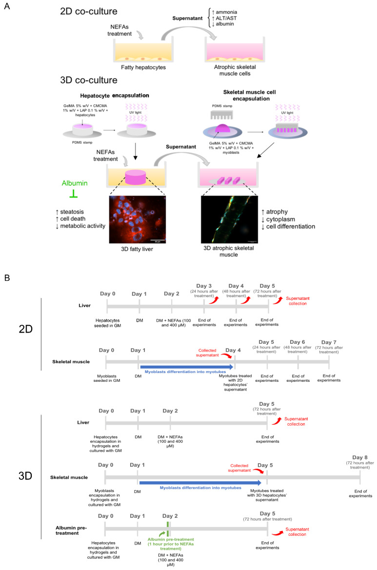
Figure 1.
(A) Schematic representation of the in vitro experimental model employed in this study. (B) Schematic representation of the timeline of the experiments performed in this study. 2D: two-dimensional; NEFAs: non-esterified fatty acids; ALT: alanine aminotransferase; AST: aspartate aminotransferase; 3D: three-dimensional; GelMA: gelatin methacryloyl; CMCMA: carboxymethyl cellulose methacrylate; LAP: lithium phenyl(2,4,6-trimethylbenzoyl)phosphonate; UV: ultraviolet; PDMS: polydimethylsiloxane; GM: growth media; DM: differentiation media.

