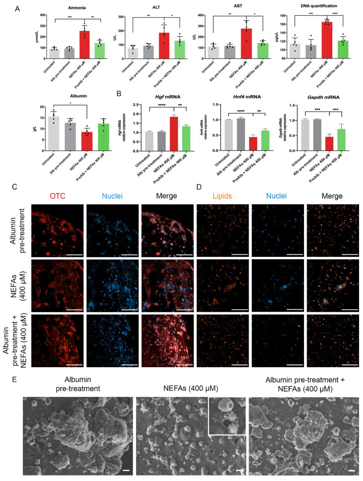
Figure 6.
Albumin pre-treatment protect 3D hepatocytes upon challenge with NEFAs. (A) Biochemical analyses of the supernatant of 3D AML12 cells pre-treated with albumin and challenged with NEFAs for 72 h assessed by clinical standard procedures. (B) Real-time qPCR of hepatocyte markers. The untreated condition was used as calibrator. (C) Confocal microscopy of a hepatocyte’s functionality marker of 3D AML12 cells pre-treated with albumin and challenged with NEFAs for 72 h. Scale bar = 200 µm. (D) Confocal images of lipids accumulation of 3D AML12 cells pre-treated with albumin and challenged with NEFAs for 72 h in NEFAs treated hepatocytes assessed by AdipoRed™ assay. Scale bar = 200 µm. (E) Ultrastructural assessment of AML12 cells phenotype by scanning electron microscopy. Scale bar = 10 µm. The results are expressed as mean values ± SEM and compared using one-way analysis of variance followed by post hoc tests when appropriate. * p < 0.05; ** p < 0.01; *** p < 0.001; **** p < 0.0001. NEFAs: non-esterified fatty acids; ALT: Alanine Aminotransferase; AST: Aspartate Aminotransferase; Hgf: hepatocyte growth factor; Hnf4a: hepatocyte nuclear factor 4 alpha; Gapdh: glyceraldehyde 3-phosphate dehydrogenase; OTC: ornithine transcarbamylase.

