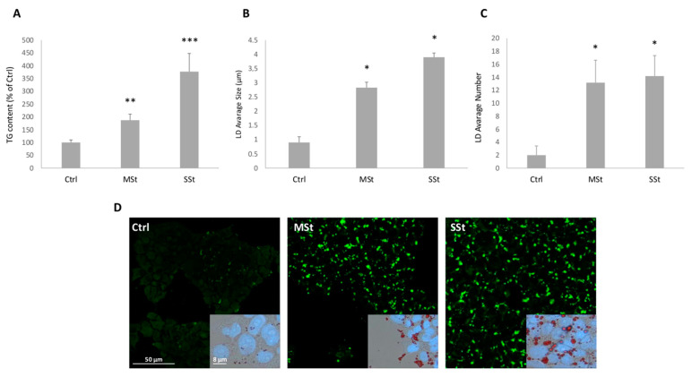
Figure 2.
Lipid accumulation in moderate and severe steatosis models. For FaO cells incubated in the absence (Ctrl) or in the presence of moderate in vitro steatosis (MSt), and severe steatosis (SSt) we show: (A) TG content expressed as percent TG content relative to controls, normalized for proteins determined with Bradford assay. (B,C) Average size of LDs. and number of LDs/cell. Values are mean ± S.D. from at least three independent experiments. Statistical significance between groups was assessed by ANOVA followed by Tukey’s test. Symbols: Ctrl vs. all treatments * p ≤ 0.05. (D) Microphotographs of cells stained with BODIPI 493/503 (magnification 20×; Bar: 50µm) and microphotographs of cells stained simultaneously with ORO and DAPI (magnification 40×; Bar: 8µm) were captured. For microscopy analyses a Leica DMRB light microscope equipped with a Leica CCD camera DFC420C was employed. Symbols: Ctrl vs. all treatments * p ≤ 0.05, ** p ≤ 0.01, *** p ≤ 0.001.

