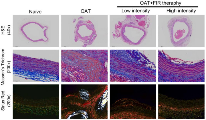
Figure 1.
FIR therapy reduced allograft vasculopathy in OAT-recipient ACI/Nkyo rats. (upper column) Thoracic aortas from donor PVG/Seac rats stained with hematoxylin and eosin. The arrows indicate internal elastic lamina and arrowheads indicate calcified lesions. The images are 40× magnified. (middle column) The integrity of collagen fibers of thoracic aorta cross-sections was observed using Masson’s trichome staining. (lower column) Histopathological features and collagen accumulation of thoracic aorta cross-sections were observed using picrosirius red staining. The slides were observed via light microscopy and polarized light microscopy, respectively (200× magnification).

