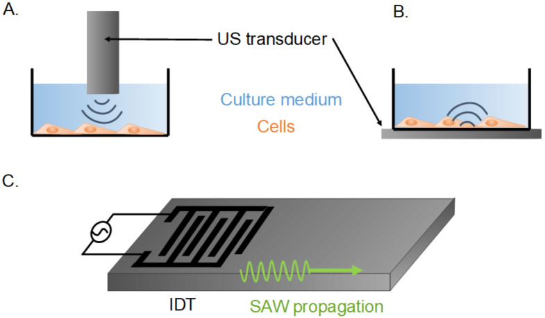
Figure 1.
Schematic view of cell-stimulation systems of low- or high-intensity ultrasound stimulation. (A): Cells stimulated mainly by the shear flow induced by a US transducer immersed in the culture well. (B): Cells stimulated mainly by the mechanical vibrations of the culture well. US stimulated by the US transducer under it. (C): Piezo-electric system with an interdigital transducer (IDT) inducing surface acoustic waves (SAW).

