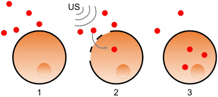
Figure 4.
Schematic description of gene or protein transfection (schematized in red dot) into a cell (schematized in orange, with its nucleus in darker orange). The elements to be transferred are in the extracellular medium (1). The cell membrane is disrupted by US (schematized by the grey waves) (2). The cell membrane closes again after integration of the transfected elements (3).

