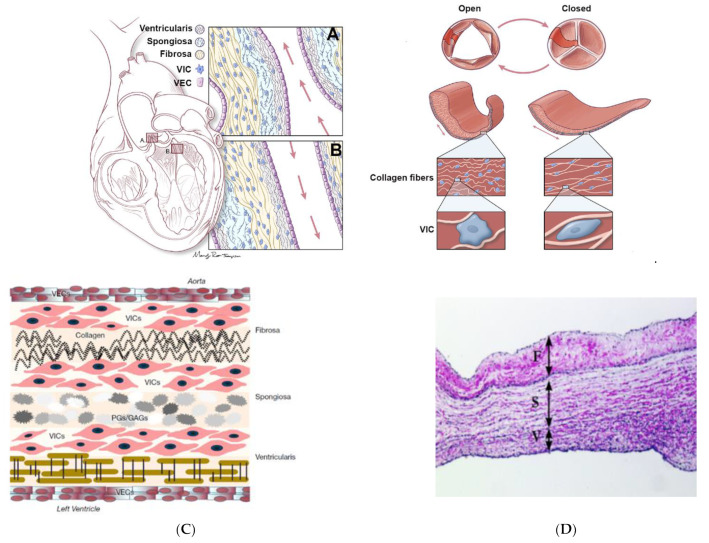
Figure 7.
Representation of aortic and mitral valve structures. (A) Aortic valve and (B) mitral valve, with the three ECM layers: ventricularis (EL), spongiosa (PG-GAG) and fibrosa (COL); the blood flow is indicated by red arrows (ventricularis closest to blood flow); valve endothelial cells (VECs, purple) and valve interstitial cells (VICs, blue). (Right) Representation of the aortic valve indicating coordinated rearrangement of the ECM fibers, and elongation of the VICs during systole (open) and diastole (closed). Reprinted with permission from ref. [111]. Copyright 2020, MDPI. (C) Detailed heart valve structure: the three inner layers (ventricularis, spongiosa and fibrosa) with proteoglycans (PG), glycosaminoglycans (GAG), collagen type I and type III, elastin and VICs and the outer layer formed by VECs. Reprinted with permission from ref. [105]. Copyright 2015, Cambridge University Pres. (D) Tissue image of trilayered structure of an aortic leaflet in sheep. The three layers consist of fibrosa (F), spongiosa (S) and ventricularis (V). Reprinted with permission from ref. [112]. Copyright 2015, SciDoc Publishers.

