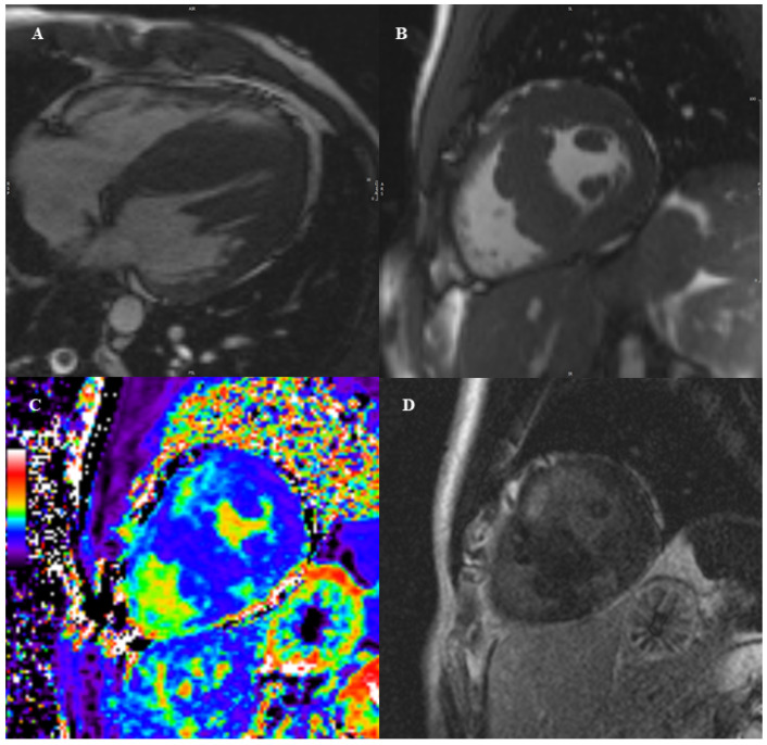
Figure 2.
13 year-old boy with positive family history for HCM. The CMR examination showed an asymmetric hypertrophy with prevalent involvement of the interventricular septum with a maximal wall thickness of 34 mm, and hypertrophy of papillary muscles (panel (A,B)), which was highly suggestive for HCM. To implement diagnosis and risk stratification, we then performed tissue characterization sequences. We added parametric mapping sequences before and after contrast injection. As shown in panel (C), the calculated extracellular volume in the mid anterior and posterior septal wall was elevated (38%), as a sign of diffuse fibrosis. In the same segments late gadolinium enhancement was also evident with a patchy, intramyocardial distribution, as a sign of replacement fibrosis (panel (D).

