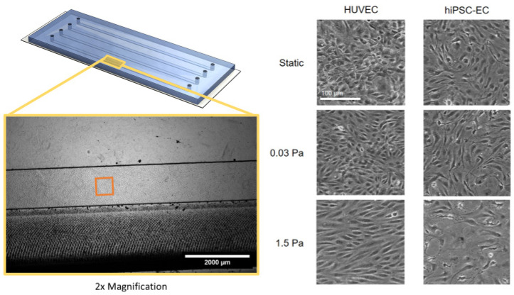
Figure 3.
Endothelial cell morphology after 24 h of exposure to specific shear stress in a microfluidic chip channel. HUVEC (left) or hiPSC-EC (right) were cultured in microfluidic chips until confluent and were subsequently left under static conditions (top), treated with flow conditions causing low shear stress (middle) or high shear stress (bottom). Differences in morphology are shown by representative phase-contrast microscopy images taken after 24 h of culturing under the respective conditions. The scale bar is 100 μm.

