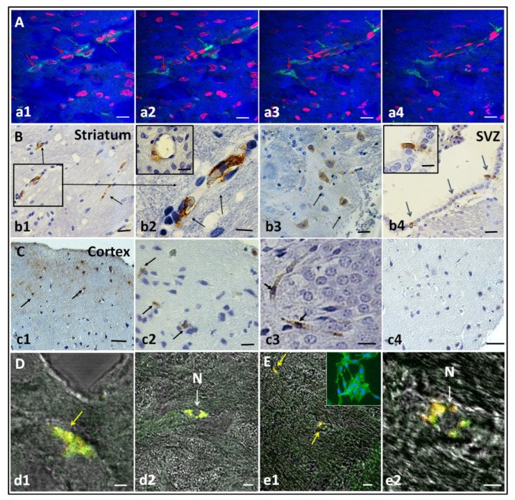
Figure 4.
Engraftment of hIDPSCs four days after IV administration in the striatum, cortex and subventricular zone (SVZ). (A) Images show hIDPSC (within striatum) stained with Vybrant (green arrows) and nuclei stained with propidium iodide (red arrows). Analysis performed at day 9 (D9). (B,C) Immunohistochemistry demonstrates positive immunostaining for the anti-hNu antibody (black arrows) observed within the striatum (b1–b3), SVZ (blue arrows) (b4) and cortex (c1–c3). B2 and B4 show cells immunolabeled with the anti-hNu antibody in the striatal perivascular area and subventricular zone, respectively. Negative control shows the absence of unspecific labeling of secondary antibody control for anti-human nuclei (c4). Analysis performed at day 9 (D9). Immunofluorescence assay (in confocal microscopy) showing the expression of CD73 (D) and CD105 in the rat brain (E). Yellow arrows demonstrate cells expressing CD73-positive (d1) and CD105-positive (e1) and, white arrows, cells C73 (d2) and CD105 in smallest magnification (e2). These cells exhibits an usual MSC phenotype (N). Analysis performed at day 35 (D35). Total magnification: 5× (a1–a4,b1,b4,c1,e1), 10× (c4,d2), 20× (c2) and 40× (b2,b3,c3,d1,e2). Scale bars: 5 µm (a1–a4,b2,b4,c2–c4,d1,d2,e1,e2), 10 μm (b3).

