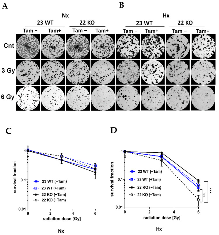
Figure 7.
Survival of mHDC in response to irradiation with up to 6 Gy in normoxia (NX) or hypoxia (HX) as determined by colony formation assay (CFA). Hypoxic cells were cultured at 1% O2 in rat collagen coated 6-well dishes. After IR, the cells designated HX were placed in hypoxia for 10 days. The cultures were fixed, stained with Coomassie and colonies of at least 50 cells were counted. Photomicrographs in (A,B) show representative pictures of colony formation upon treatment with IR without or with Tam treatment for Nx (A) or moderately hypoxic conditions (B). Survival curves shown in (C,D) depict quantification of colony formation upon treatment in normoxia (C) and hypoxia (D). All groups consisted of n = 6 independent cultures. Groups were compared using two-way ANOVA. (** p < 0.01; *** p < 0.001).

