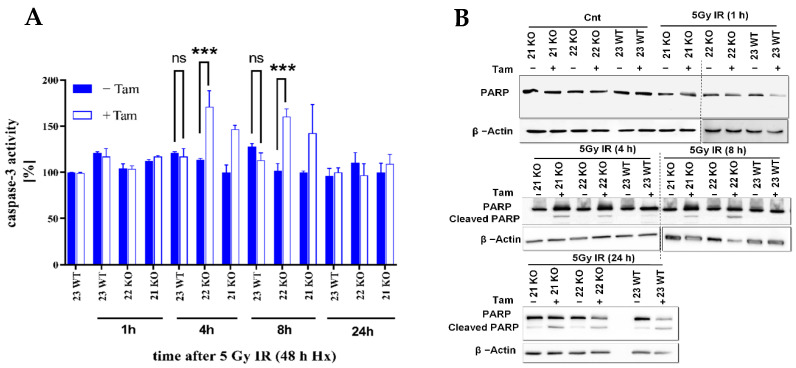
Figure 8.
Analysis of apoptosis induction in hypoxic HIF-1α deficient mHDC. (A) The activity of caspase-3 was measured in mHDC lysates. Cells were irradiated and incubated in hypoxia for the time indicated. The cells were then lysed, and 10 µg protein were incubated with Ac-DEVD-amido-4-methylcoumarine. Generation of the fluorescent product was monitored at 430 nm. Caspase-3 activity data showed representative experiments with n = 6, each experiment was repeated three times. Graphs show mean ± SD, groups were compared using two-way ANOVA, *** p < 0.001, ns p > 0.05. (B) Western blot with a PARP-1 antibody which detects full-length PARP and a cleaved species with a lower molecular weight indicative of PARP cleavage, a signature of apoptosis. The cells were incubated in hypoxia for 48 h, then irradiated with 5 Gy, and transferred back to hypoxia for the time indicated. The cells were then lysed and subjected to SDS-PAGE and Western blotting. The data show a representative Western blot from three independent experiments.

