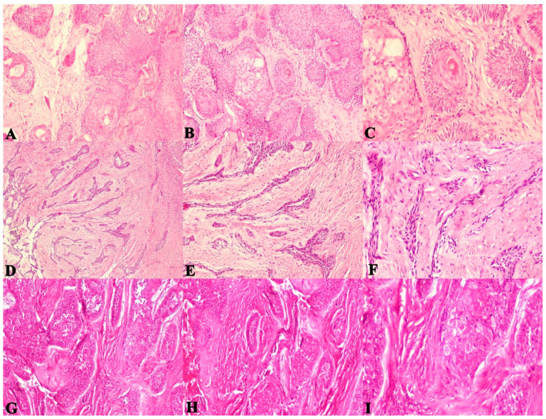
Figure 6.
Photomicrographs of haematoxylin and eosin stain. Acanthomatous ameloblastoma exhibiting extensive hyalinization between follicles, (A). 40×, (B). 100× and (C). 400× Desmoplastic ameloblastoma exhibiting thin compressed strands due to desmoplastic changes and hyalinization, (D). 40×, (E). 100× and (F). 400× Granular cell ameloblastoma exhibiting extensive hyalinization between follicles filled with large granular cells, (G). 40×, (H). 100× and (I). 400×.

