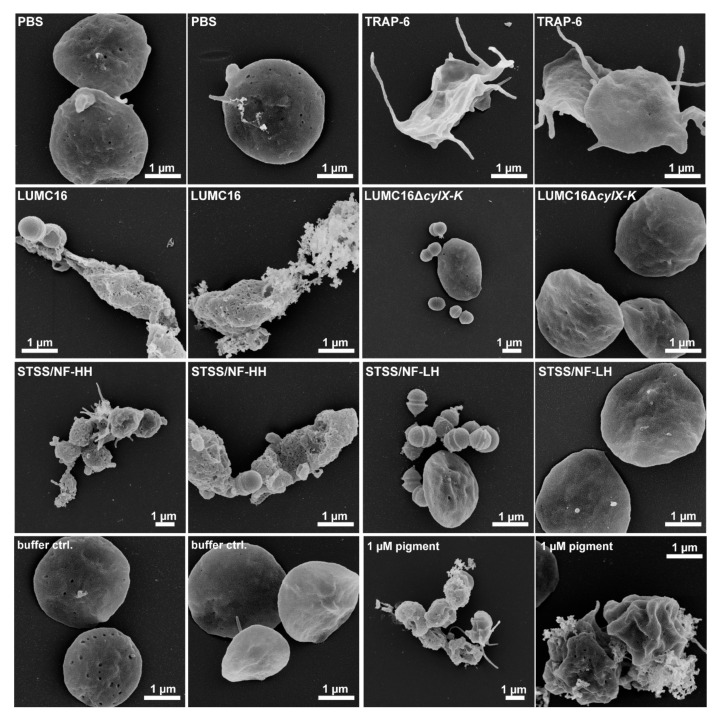
Figure 4.
Platelet morphology in response to GBS infection or pigment stimulation. Washed human platelets were infected with indicated GBS strains or stimulated with pigment (1 µM) for 120 min and were visualized via FESEM. PBS and TRAP-6 were used as negative and activation controls, respectively. Representative images from three independent experiments are shown (n = 3).

