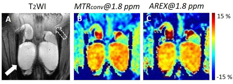
Figure 1.
Representative in vivo Cr-CEST images of control mouse testes. (A) Anatomical T2WI image. The white arrow shows the testis. The white dotted arrow shows the testicular epithelium. (B) MTR asymmetry maps at 1.8 ppm of MTRconv. (C) MTR asymmetry maps at 1.8 ppm of AREX. MTR, magnetization transfer ratio; MTRconv, conventional analysis metric magnetization transfer ratio; AREX, apparent exchange-dependent relaxation; and Cr-CEST, creatine chemical exchange saturation transfer.

