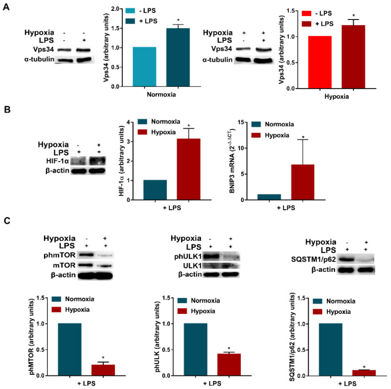
Figure 4.
LPS enhances Vps34 expression and promotes autophagy in hypoxic DCs. (A) Vps34 protein levels in DCs exposed to normoxia and hypoxia, in the presence or not of LPS, for 24 h. (B) HIF-1α protein levels, as determined by Western blotting and BNIP3 mRNAs as determined by RT-qPCR analysis, respectively, in DCs stimulated with LPS and exposed to normoxia and hypoxia for 24 h. (C) phmTOR, mTOR, phULK1, ULK1 and SQSTM1/p62 protein levels by Western blotting. All blots are representative of at least three independent experiments. β-actin was used as loading control and as housekeeping gene for RT-qPCR analysis. Asterisk indicates statistically significant differences (p ≤ 0.05; n = 3).

