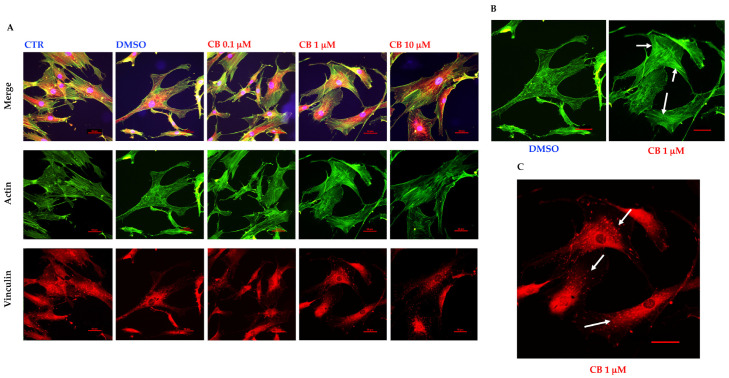
Figure 7.
Immunofluorescence of human adipose-derived stem cells’ (hASCs) cytoskeletal markers after 24 h of recovery time from the cytochalasin B (CB) treatment. (A) hASCs were immunostained with Phalloidin (green signal, specific for F-actin) and anti-vinculin (red signal) 24 h after the treatment wash-out. Acquisitions of images were made with a Nikon inverted microscope Eclipse Ti2-E and a digital sight camera DS-Qi2, through the imaging software NIS-Elements. (B) Enlarged images referring to cells previously treated for 24 h with 0.05% dimethyl sulfoxide (DMSO) (CB vehicle) or CB 1 μM, after 24 h of recovery time. White arrows indicate the bundle of actin. (C) Enlarged images referring to cells previously treated for 24 h with CB 1 μM, after 24 h of recovery time. White arrows indicate some focal adhesions. Scale bars: 50 μm.

