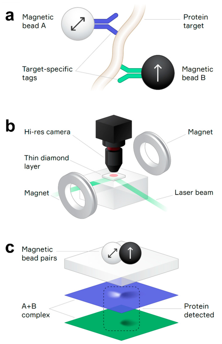
Figure 1.
(a) Immunocomplex containing two magnetic beads covalently bonded to antibody tags specific to different epitopes of the target protein analyte. Arrows indicate the different magnetic properties of magnetic beads A and B, which are superparamagnetic and ferromagnetic, respectively. (b) Schematic of the quantum diamond microscope featuring a synthetic diamond chip with a thin layer of NV centers, a laser beam to excite NV center fluorescence, a high-resolution objective lens to collect the fluorescence onto a camera, and an electromagnet to apply different bias magnetic fields to the magnetic beads in the sample. (c) An immunocomplex containing both magnetic bead A and bead B is identified by overlapping magnetic dipole signals (light + dark lobes) in the separated bead A and bead B detection channels (blue and green images, respectively).

