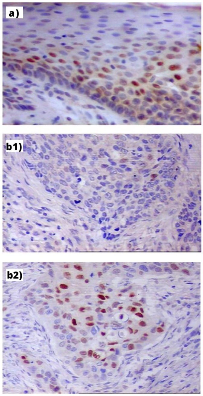
Figure 1.
Immunohistochemical expression of Cyclin D1 in: (a) PLL—visible mostly in the nuclei of basal layer cells of hyperplastic epithelium (index: 32%, orig. magn. 200×); (b) LC: (b1) in the GG homozygote patient—visible in single cancer cells (index: 5%, orig. magn. 200×); (b2) in the AA homozygote patient visible as small cellular granules in nuclei of cells (index: 41%, orig. magn. 200×).

