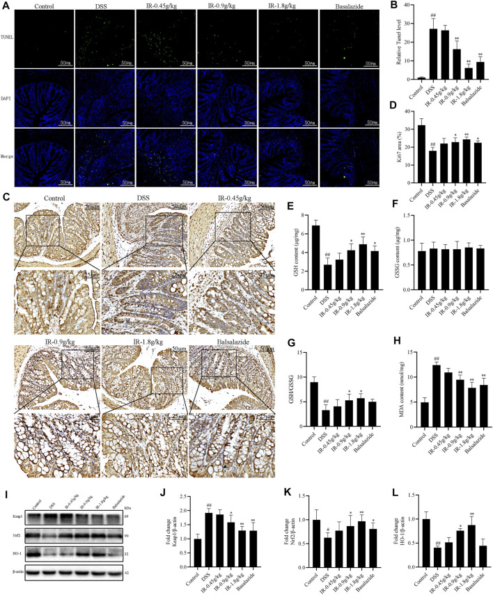
FIGURE 4.
IR alleviates the apoptosis and oxidative stress of colon tissue in UC mice. (A) Colon cell apoptosis detected by Tunel staining. (B) Positive area statistics of Tunel. (C) Colonic epithelial cell proliferation detected by Ki67 staining. (D) Histochemical positive area statistics of Ki67. (E,F) The content of glutathione (GSH) and oxidized glutathione (GSSG). (G) The ratio of GSH/GSSG. (H) The content of malondialdehyde (MDA). (I) The protein levels of Keap1, Nrf2, and HO-1 in colon detected by western blotting. (J-L) The gray intensity analysis of Keap1, Nrf2, and HO-1 proteins, respectively. All data were compared using one-way ANOVA, and p-values reflected differences between experimental groups (n = 3).

