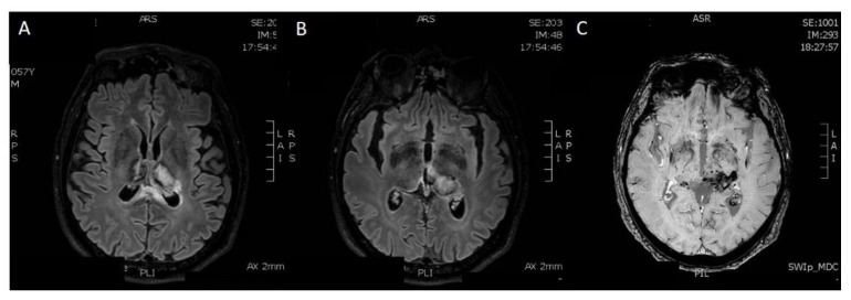
Figure 6.
Five months later, he underwent a brain MRI exam. (A,B) Axial FLAIR-weighted sequences show reduction in the diameter of haemorrhagic focus and reduction in perilesional oedema in both thalami. (C) In the Susceptibility-Weighted Images, old hematic deposits can be found in both thalami, particularly on the left. Therefore, the exam showed a tendency to encephalomalacic transformation of the ischemic regions and improved sinus rectum patency, with some wall irregularities. The patient continued treatment with Enoxaparin.

