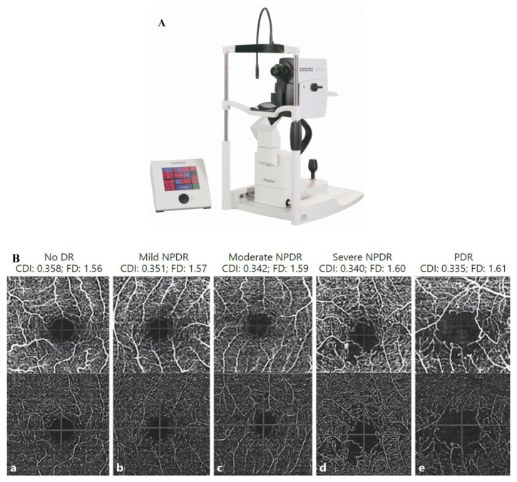
Figure 7.
(A) HRA + OCT imaging with a Spectralis HRA+ device Binarized optical coherence tomography pictures with varying degrees of DR severity, as well as non-segmented angiograms, are shown in (B). (a) There is no DR. (b) Mild NPDR if any. (c) NPDR of a moderate level. (d) A very bad case of NPDR. (e) It is a PDR and it is important to note that the following CDI and FD values are the same: CDI is 0.358 and FD is 1.56, CDI is 0.351 and FD is 1.57, CDI is 0.342 and FD is 1.59, CDI is 0.340 and FD is 1.60 and CDI is 0.335 and FD is 1.61. Nonproliferative diabetes retinopathy is referred to as NPDR, while proliferative diabetic retinopathy is referred to as PDR.

