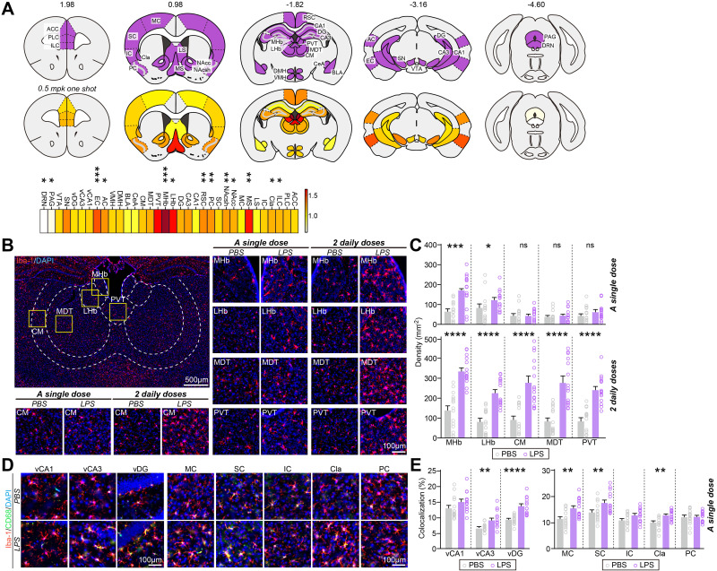
Figure 1.
Mild, LPS-induced inflammation produces a spatially patterned microglial activation throughout the adult mouse brain. (A) Heat maps of fold changes in microglia number in each anatomical region induced by single i.p. LPS injection. Top: Purple represents brain regions examined; bottom: color scale indicates fold changes in microglia number ranging from 0 to 1.64; fold change in MHb is not included (2.61) (*p < 0.05, **p < 0.01, ***p < 0.001). (B) Representative images of MHb, LHb, CM, MDT or PVT brain regions and immunostaining for the microglial marker Iba-1 in mice injected with saline or i.p.-administered a single or 2 daily doses of LPS. (C) Quantification of the density of Iba-1+ cells. Data are means ± SEMs (n = 14~15 sections from 5 mice; *p < 0.05, ***p < 0.001, ****p < 0.0001; Mann–Whitney U-test). (D) Representative images of vCA1, vCA3, vDG, MC, SC, IC, Cla or PC brain regions and immunostaining for the microglial marker Iba-1 and CD68 in mice injected with saline or i.p.-administered a single dose of LPS. Scale bar: 25 µm (applies to all images) (E) Quantification of the colocalization percentage of Iba-1+/CD68+ cells. Data are means ± SEMs (n = 14~15 sections from 5 mice; **p < 0.01, ****p < 0.0001; Mann–Whitney U-test).
Abbreviations: ACC, anterior cingulate cortex; AC, auditory cortex; BLA, basolateral nucleus of amygdala; CA1, cornu ammonis 1; CA3, cornu ammonis 3; CeA, central nucleus of amygdala; Cla, claustrum; CM, central nucleus of thalamus; DG, dentate gyrus; DMH, dorsomedial hypothalamus; DRN, dorsal raphe nucleus; EC, entorhinal cortex; IC, insula cortex; ILC, infralimbic cortex; LHb, lateral habenula; LS, lateral septum; MDT, mediodorsal nucleus of thalamus; MHb, medial habenula; MC, motor cortex; MS, medial septum; NAcc, nucleus accumbens core; NAcsh, nucleus accumbens shell; PAG, periaqueductal area; PC, piriform cortex; PLC, prelimbic cortex; PVT, paraventricular nucleus of thalamus; RSC, retrosplenial cortex; SC, somatosensory cortex; SN, substantia nigra; vCA1, ventral CA1; vCA3, ventral CA3; vDG, ventral DG; VMH, ventromedial hypothalamus; and VTA, ventral tegmental area.

