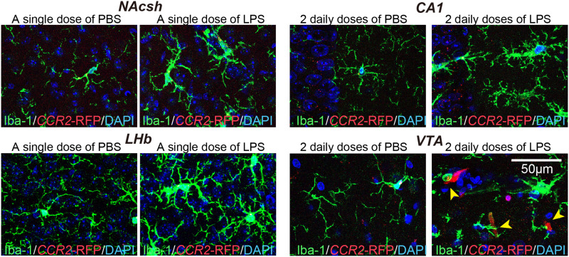
Figure 2.
Mild, LPS-induced inflammation does not cause the infiltration of peripheral immune cells. Representative images showing the number of CCR2-RFP+ cells and immunostaining for the microglial marker Iba-1 (green) in the indicated brain regions of CCR2-RFP mice injected with saline or i.p.-administered a single or 2 daily doses of LPS. Yellow arrow heads indicate CCR2-RFP+ cells.
Abbreviations: CA1, cornu ammonis 1; LHb, lateral habenula; NAcsh, nucleus accumbens shell; and VTA, ventral tegmental area.

