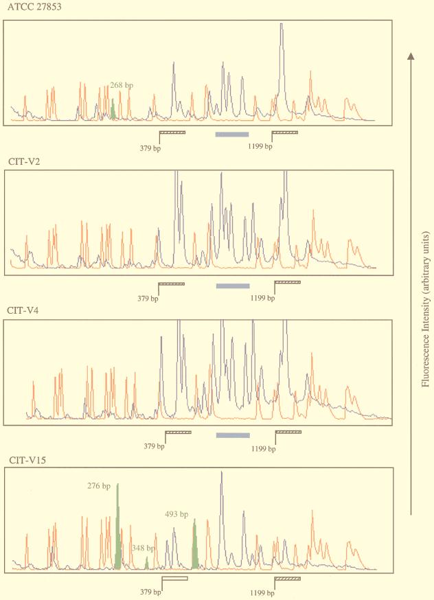
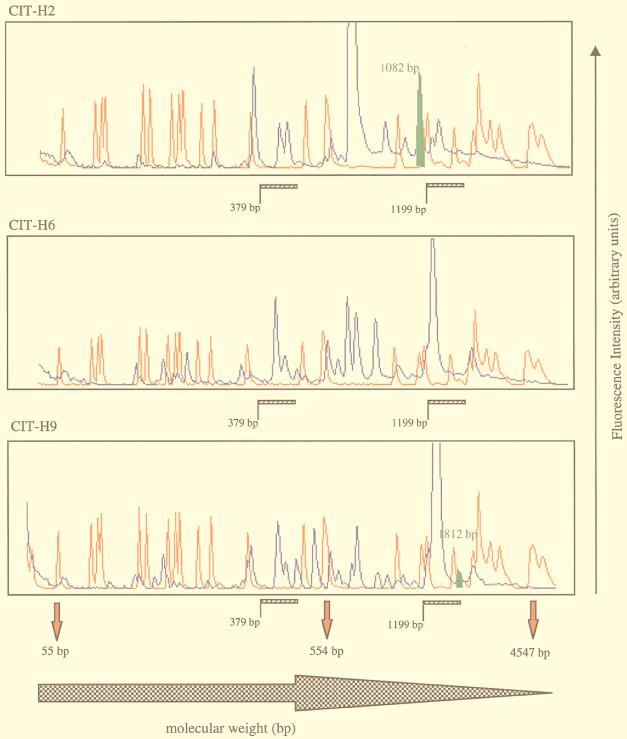
FIG. 3.
GeneScan traces of Pseudomonas isolates. Blue peaks represent 6-carboxyfluorescine-labelled RW3A-generated DNA fragments which were sized by direct comparison with the 6-carboxy-X-rhodamine-labelled internal standards (red peaks). The size range is indicated by the solid red arrowheads below the final trace. Regions of interest within the Pseudomonas genome are indicated by hatched and open bars and a solid blue bar for some isolates. Polymorphic DNA fragments are denoted by the solid green peak with corresponding molecular lengths.


