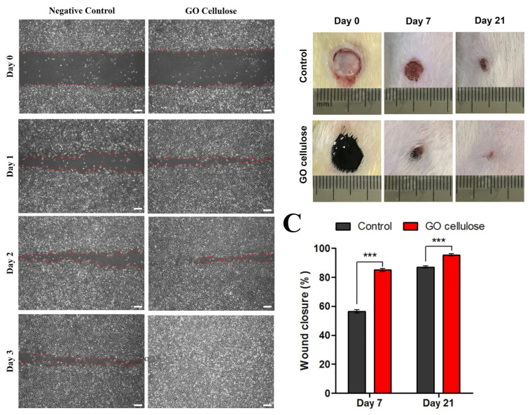
Figure 9.
(A) Cell migration with and without GO–cellulose nanocomposite; red-dotted lines represent the wound edges, scale bar = 200 μm; (B) in vivo evaluation of the skin wounds of rats with and without GO–cellulose nanocomposite for post-wound induction on days 0, 7, and 21; and (C) the percentage of wound closure: significant differences were evaluated using one-way ANOVA, where *** p < 0.0001. Reprinted with permission from Ref. [230]. 2021, Elsevier.

