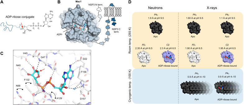
Fig. 1. The NSP3 macrodomain (Mac1) reverses mono-ADP ribosylation.
(A) Chemical structure of ADPr conjugated to a glutamate residue. The C1″ covalent attachment point is shown with a red arrow. (B) Cartoon of the multidomain NSP3 showing Mac1 with ADPr bound in the active site (PDB code 7KQP). (C) Structure of ADPr bound in the macrodomain active site (PDB code 7KQP) with the changes in protein structure upon ADPr binding indicated with black arrows. The C1″ covalent attachment point is shown with a red arrow. (D) Summary of the crystal structures reported in this work.

