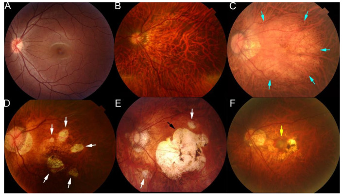
Figure 2.
Representative fundus photographs showing the different types of lesions of maculopathy in eyes with pathologic myopia. (A) Normal fundus image. (B) Tessellated fundus. (C) Diffuse atrophy around optic disc and posterior fundus (blue arrows). (D) Patchy atrophy fundus (white arrows). (E) A fundus image from a left eye with macular atrophy at the center of posterior fundus (black arrow). Patchy atrophy (white arrow) as well as diffuse atrophy background can also be seen. (F) Fundus image with myopic choroidal neovascularization at the center of fundus (yellow arrow). Reprinted from Deep Learning Approach for Automated Detection of Myopic Maculopathy and Pathologic Myopia in Fundus Images, Vol 5, Pages No. 1235–1244, Copyright (2021), with permission from Elsevier.

