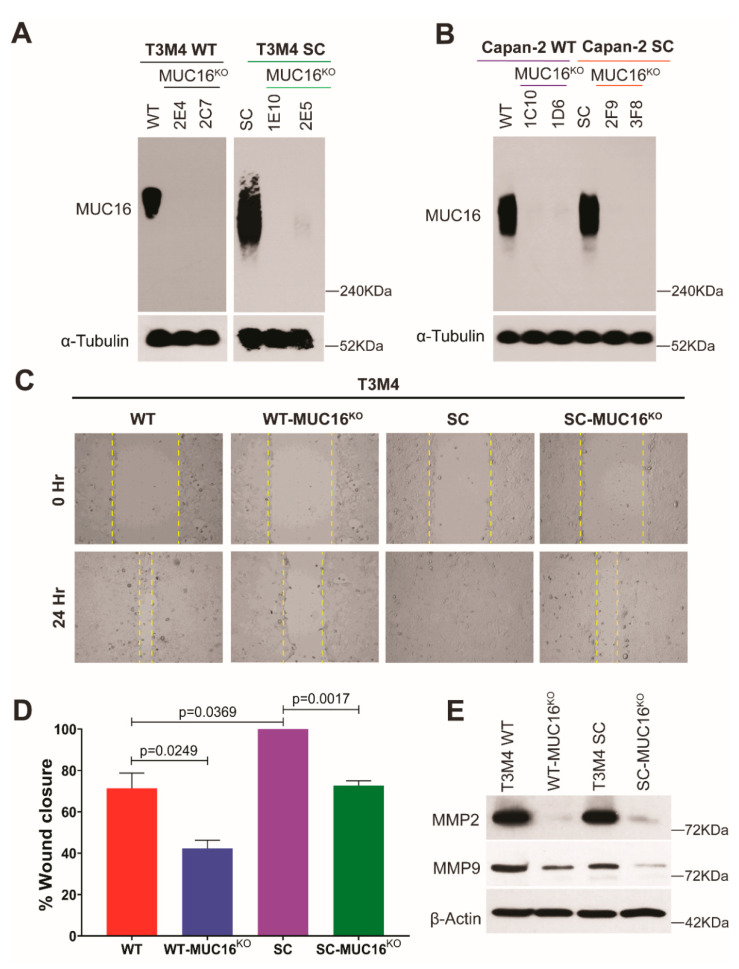
Figure 1.
MUC16 knockout causes a decrease in PDAC cell migration. Western blot confirmation of CRISPR-Cas9-mediated genetic deletion of MUC16 in (A) T3M4 WT and SC cells; and (B) Capan-2 WT and SC cells; loading control: α-tubulin; (C) wound healing assay with T3M4 WT, WT-MUC16KO, SC, SC-MUC16KO cells; images captured at 0 h and 24 h; (D) quantification of the percentage of wound closure, as described previously [14]; p < 0.05 is considered statistically significant; (E) Western blot analysis of MMP2 and MMP9 in T3M4 WT, WT-MUC16KO, SC, SC-MUC16KO cells; loading control: β-actin.

