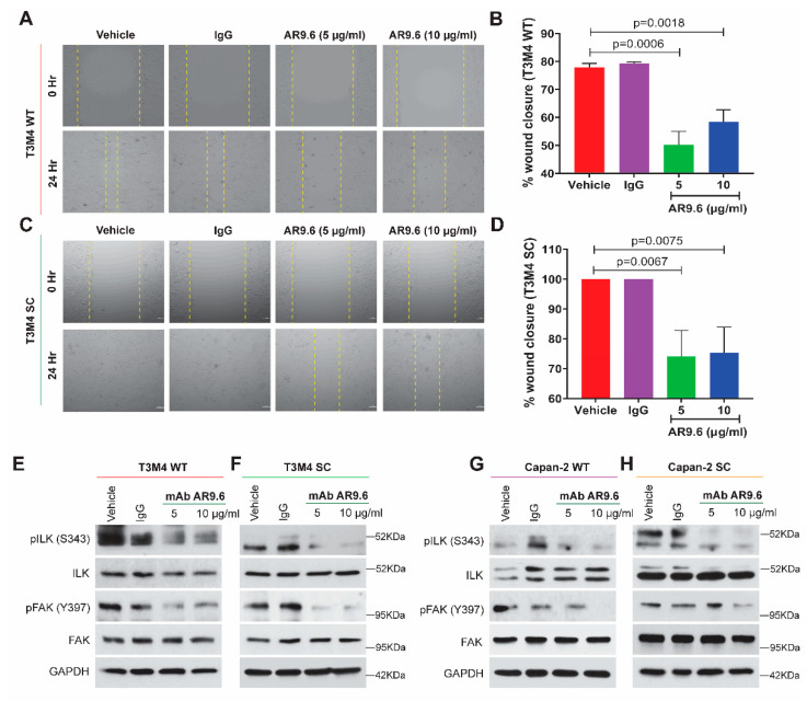
Figure 5.
Treatment with anti-MUC16 AR9.6 antibody alleviates PDAC cell migration via the ILK-FAK axis. Cells were treated with anti-MUC16 AR9.6 antibody (5 µg/mL and 10 µg/mL); controls: untreated (complete media) and isotype-control (5 µg/mL murine IgG) for all of the following: wound healing assay with (A) T3M4 WT cells; (B) quantification of percent wound closure in T3M4 WT cells; (C) T3M4 SC cells; (D) quantification of percent wound closure in T3M4 SC cells; p < 0.05 is considered statistically significant. Western blot analysis for pILK (S343), total ILK, pFAK (Y397), total FAK in (E) T3M4 WT; (F) T3M4 SC; (G) Capan-2 WT; (H) Capan-2 SC cells; loading control: GAPDH.

