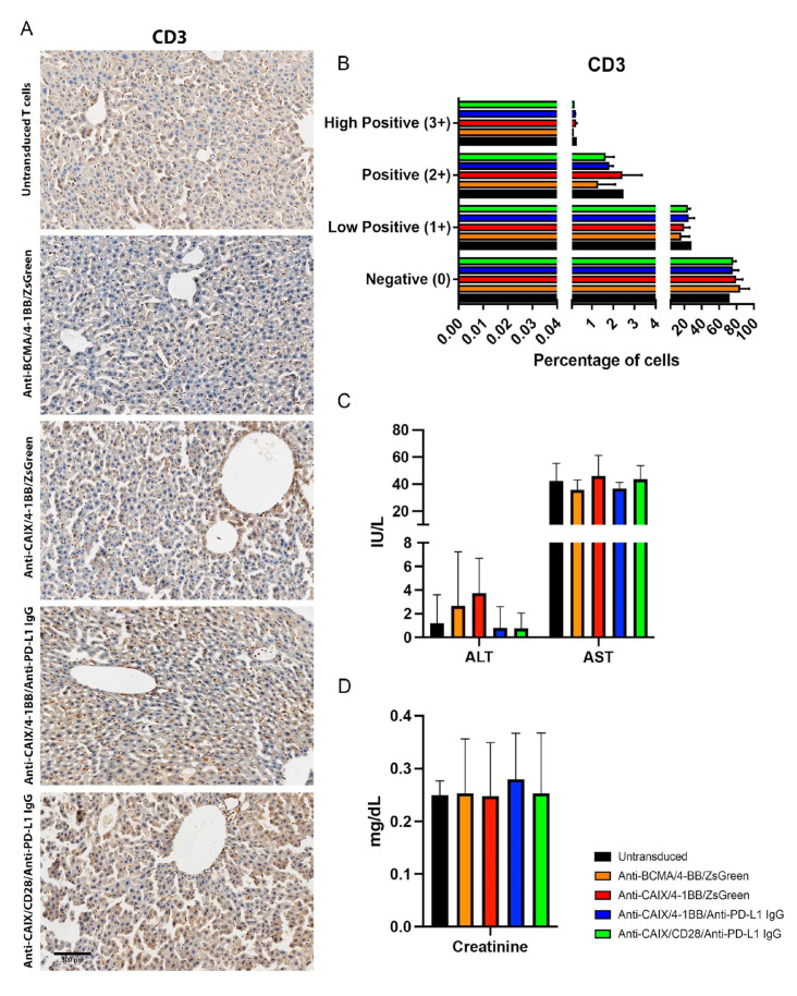
Figure 5.
Evaluation of liver infiltrating CD3+ cells and plasmatic quantification of enzymes indicative of hepatic and renal injury in mice after chimeric antigen receptor (CAR) T cells therapy. (A) Immunohistochemistry (IHC) for CD3+ T cells detection in the liver. The scale bars represent the magnification of the images of each column [300 μm (20×)]. (B) CD3 Quantification. The quantification of the IHC images was performed using the IHC Profiler Plugin of ImageJ Software [22]. The percentages of negative (0), low positive (1+), positive (2+), or high positive (3+) cells were shown. (C) Alanine transaminase (ALT), aspartate transaminase (AST), and (D) creatinine plasmatic dosage in mice. The dosages were performed in the mice plasma by the spectrophotometric method (Cobas 8000, Roche/Hitachi) one month after the beginning of the therapy with two injections of 3 × 10⁷ CAR T cells/kg with fifteen days of interval between them.

