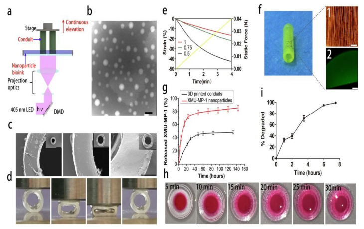
Figure 10.
(a) Schematic diagram of a hydrogel catheter prepared with the DLP 3D printing method; (b) TEM image of XMUMP−1 nanoparticles; (c) SEM images of 3D-printed conduits with different size. (Scale bar at the low magnification = 1 mm, scale bar at a high magnification = 200 lm). (d) The compression of the conduits with various wall thickness (0.5 mm, 0.75 mm, 1 mm) and the quantitative analysis (e). (f) The micro-structure (scale bar = 40 lm) (imaged by digital camera) and the nanoparticles distribution of the conduit (scale bar = 500 lm) (imaged by confocal microscopy). (g) In vitro release ofXMU-MP-1 from XMU-MP-1 nanoparticles and nanoparticle-enhanced conduits. (h) The diffusion of small molecule in the conduits. (i) The degradation of the conduits in collagenase solution.

