. 2022 Apr 11;11:e72805. doi: 10.7554/eLife.72805

Figure 5. Integrin ligation-dependent immune synapse (IS) formation requires myosin 2A contractility.

(A1–A3) DMSO-treated, phalloidin-stained primary B cells 15 min after engagement with a PLB containing ICAM-1 and limiting anti-IgM. (B1–B3) Same as (A1–A3) except the B cells were treated with pnBB. (C1–C3) Images of a representative, DMSO-treated primary B cell (white arrows mark actin arcs). (D1–D3) Images of a representative, pnBB-treated primary B cell. (E) Percent of cells exhibiting centralized, partially centralized, and noncentralized antigen (see Figure 5—figure supplement 1D1–D3 for representative examples of these three types of antigen distribution) (N = 126–144 cells/condition from three experiments). (F) Percent of total synaptic antigen in the cSMAC (N = 81–86 cells/condition from three experiments). (G) Antigen cluster size as a function of normalized distance from the cSMAC center (N = 113–144 cells/condition from three experiments). (H) Total synaptic antigen content (N = 56–62 cells/condition from three experiments). All panels: Airyscan images. Scale bars: 10 µm in (A1, B1, A3, B3); 5 µm in (D3).

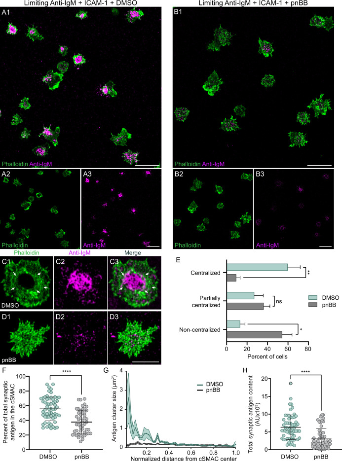
Figure 5.

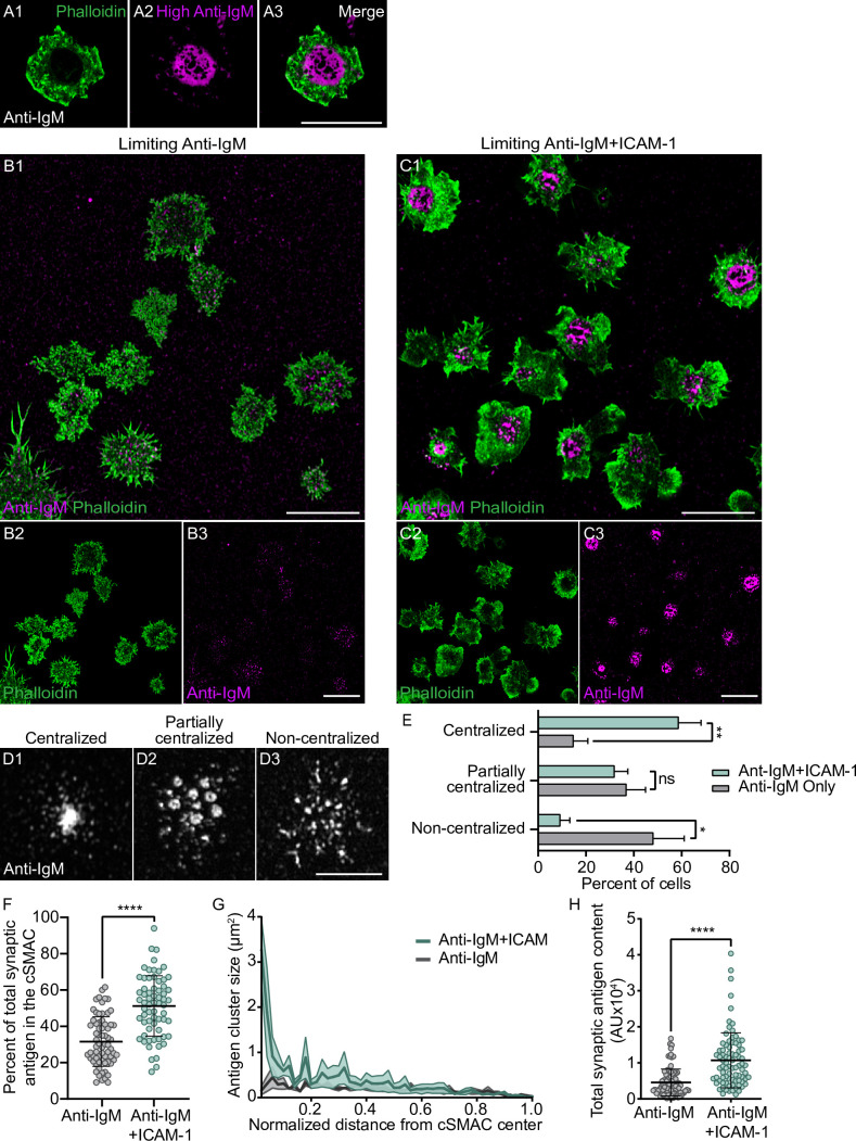
Figure 5—figure supplement 1. ICAM-1 co-stimulation promotes antigen centralization and immune synapse (IS) formation when antigen is limiting.

Figure 5—figure supplement 1.

(A1–A3) Representative, phalloidin-stained primary B cell 15 min after engagement with a PLB containing fluorescent anti-IgM at high density. (B1–B3) Phalloidin-stained primary B cells 15 min after engagement with a PLB containing a limiting amount of anti-IgM. (C1–C3) Same as (B1–B3) except the PLB also contained ICAM-1. (D1–D3) Representative images of centralized, partially centralized, and noncentralized antigen. (E) Percent of cells exhibiting the three types of antigen distribution shown in (D1–D3) (N = 151 cells/condition from four experiments). (F) Percent of total synaptic antigen in the cSMAC (N = 66–68 cells/condition from three experiments). (G) Antigen cluster size as a function of normalized distance from the cSMAC center (N = 62–69 cells/condition from three experiments). (H) Total synaptic antigen content (N = 83–87 cells/condition from three experiments). All panels: Airyscan images. Scale bars: 10 µm.
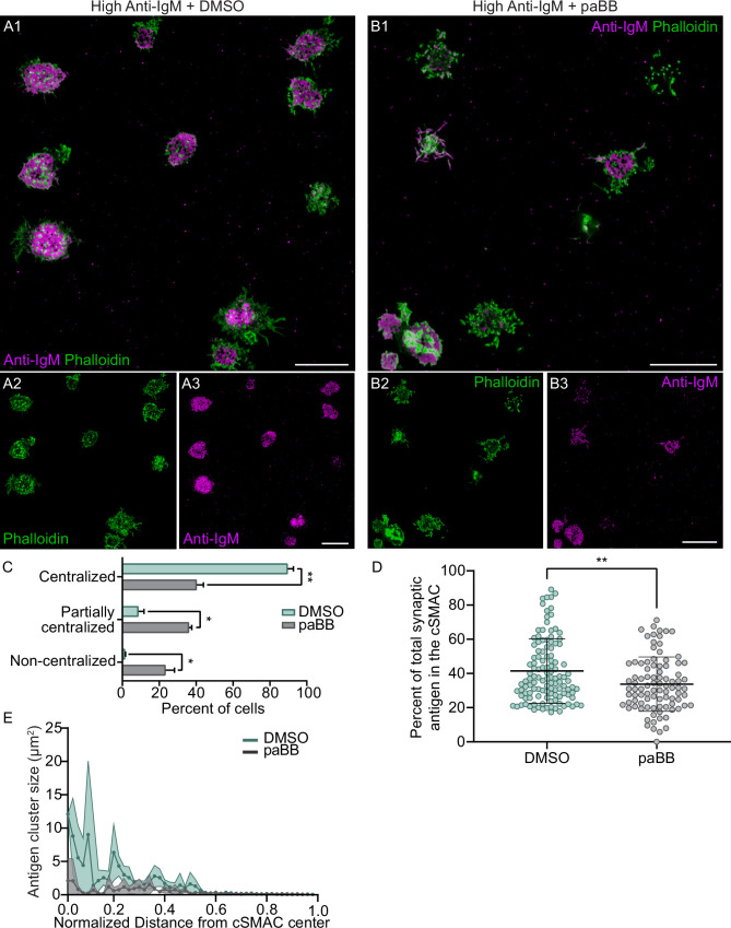
Figure 5—figure supplement 2. M2A contractility potentiates antigen centralization even when antigen density is high.

Figure 5—figure supplement 2.

(A1–A3) Phalloidin-stained, DMSO-treated primary B cells 15 min after engagement with a PLB containing high-density anti-IgM. (B1–B3) Same as (A1–A3) except the cells were treated with paBB. (C) Percent of cells exhibiting centralized, partially centralized, and noncentralized antigen (N = 137–198 cells/condition from three experiments). (D) Percent of total synaptic antigen in the cSMAC (N = 91–121 cells/condition from three experiments). (E) Antigen cluster size as a function of normalized distance from the cSMAC center (N = 86–130 cells/condition from three experiments). All panels: Airyscan images. Scale bars: 10 µm.