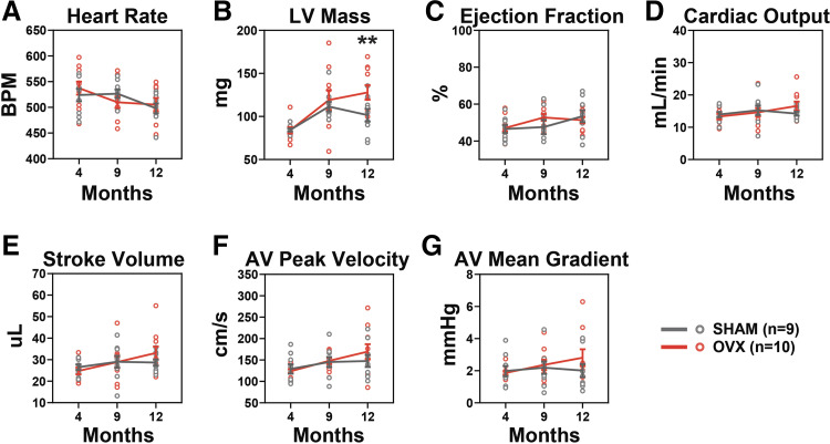
Figure 2.
OVX results in increased LV mass. M-mode and pulsed wave Doppler echocardiography were used to compare AV and LV metrics at 4, 9, and 12 mo of age. A: heart rate decreased over time but did not differ between groups. B: LV mass was significantly increased in OVX compared with sham at 12 mo. C: there was no functional change noted in either group as measured by an ejection fraction of the LV. Cardiac output (D) and stroke volume (E) did not differ between groups. F: peak velocity was increased in OVX over time but there was no group difference. G: there was no change in either group in the mean pressure gradient across the AV. Means ± SE, two-way ANOVA. Significance markers indicate group (sham vs. OVX) changes at each time point. **P < 0.01. AV, aortic valve; LV, left ventricle; OVX, ovariectomy.

