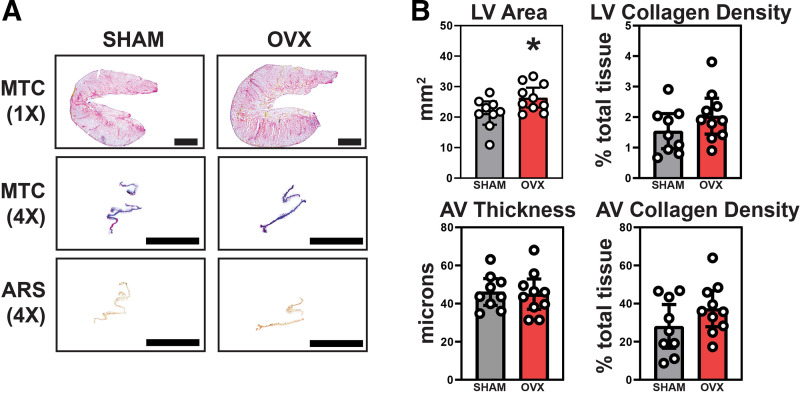
Figure 3.
LV hypertrophy was noted in OVX hearts via ex vivo analysis, but valves were absent of any significant thickening, collagen alteration, or calcification. A: Masson’s Trichrome staining was used to study gross and collagen-specific morphology in the LV (top row) and AV (middle row). Alizarin Red S staining was used to identify potential regions of calcification on the AV leaflets (bottom row). B: the LV area (top left) was significantly greater in OVX mice bolstering the LV hypertrophic phenotype noted in echocardiography. There was no significant difference in LV fibrosis (top right). Measurement of AV fibrosa-ventricularis thickness (bottom left) and collagen density (bottom right) showed no difference between groups further proving a significant AV disease phenotype secondary to OVX unlikely. A: scale bar magnitudes: 1× = 10 mm, 4× = 1 mm. B: means ± 95% CI, unpaired Student’s t test, *P < 0.05. AV, aortic valve; LV, left ventricle; OVX, ovariectomy.

