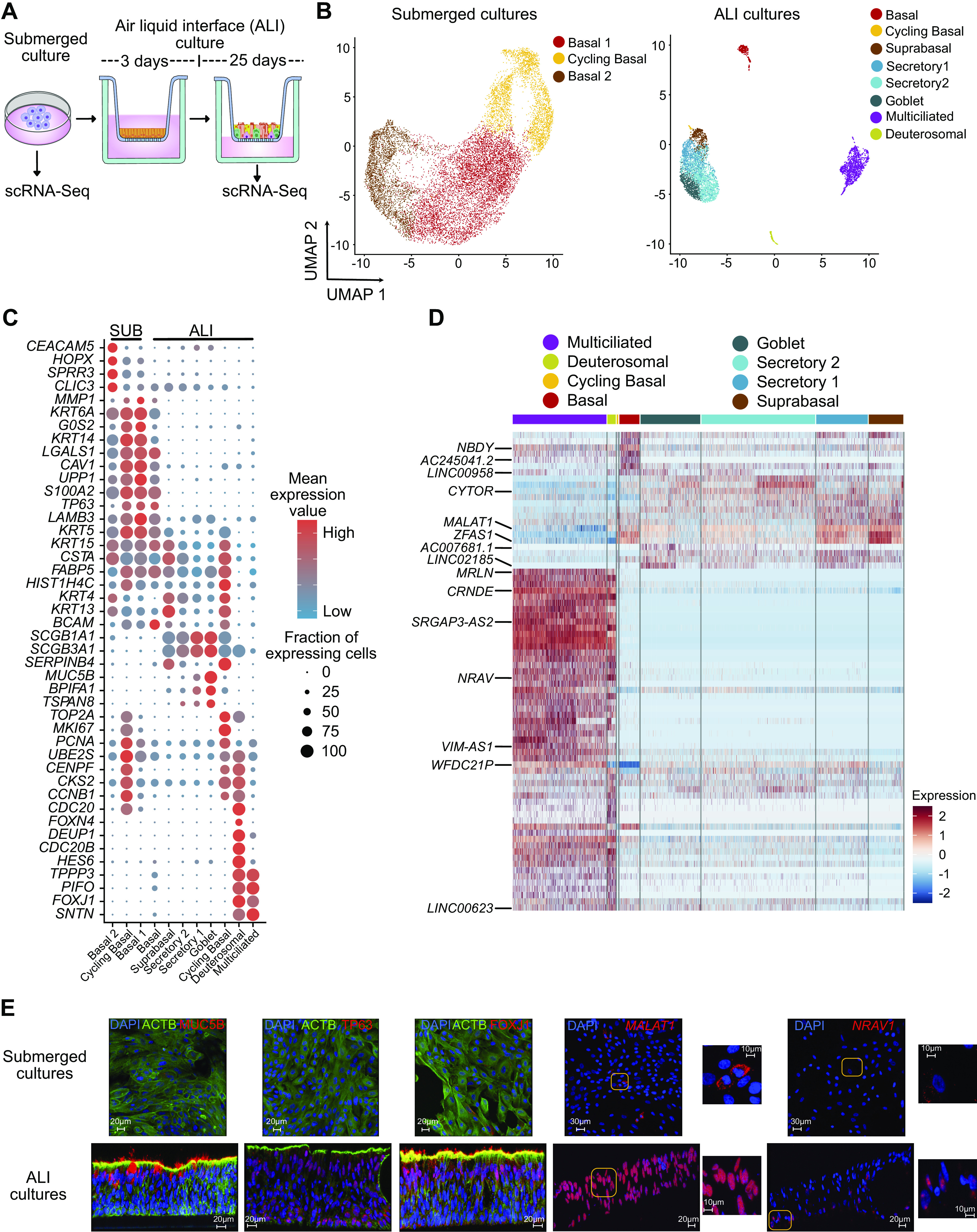
Figure 2.

Single-cell analysis reveals epithelial cell-type-specific lncRNAs. A: schematic representation of submerged and ALI cultures. Primary human bronchial epithelial cells (HBECs) were dissociated from either submerged or ALI cultures before analysis by scRNA-Seq, n = 3 donors. B: UMAP plot of the scRNA-Seq expression data highlighting the main cell clusters observed in (left) submerged cultures and (right) ALI cultures. C: expression data highlighting selected cell-specific markers for cell clusters in submerged cultures and ALI cultures. D: heatmap depicting relative expression (normalized and scaled expression) of lncRNAs in each cluster in ALI cultures. All lncRNA names and their respective expression values are available in Supplemental Table S7. E: immunofluorescence (MUC5B, TP63, and FOXJ1) and RNA in situ hybridization (MALAT1 and NRAV) demonstrating expression of selected protein and RNA molecules in submerged and ALI cultures. Scales are depicted as micrometers; n = 2 donors, representative data from one donor is shown. ALI, air-liquid interface; lncRNA, long noncoding RNA; scRNA-Seq, single-cell RNA-sequencing.
