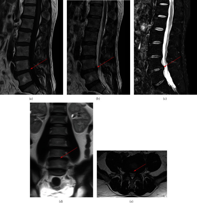
Figure 6.

MRI examination of a patient with nonspinal tuberculosis. The arrow points to the location of the lesion. (a) MRI image in the sagittal T1 sequence. (b) MRI image in the sagittal T2 sequence. (c) MRI image of T2 lipid compression sequence in sagittal position. (d) MRI image in coronal position. (e) MRI image in cross-section.
