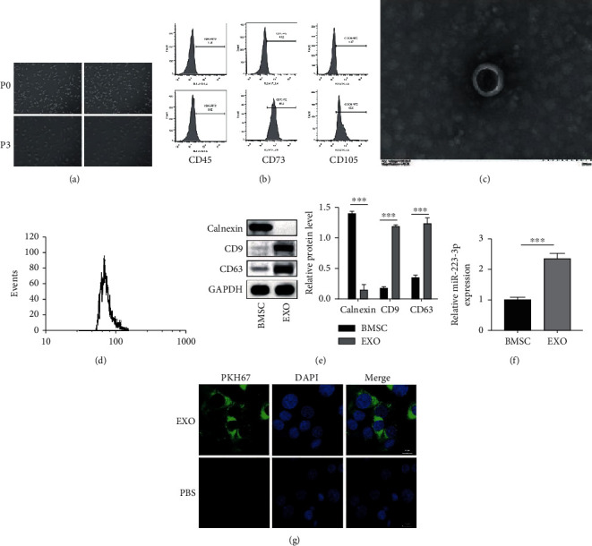
Figure 1.

Isolation of BMSCs and BMSC-derived EVs. (a) The morphology of the BMSCs was examined under a light microscope. (b) Flow cytometry was used for identifying the BMSC markers. (c) The morphology of the EVs was observed using TEM. (d) The distribution of the exosome diameters was analyzed. (e) The exosome-specific markers were detected using the western blot analysis. (f) The enrichment of miR-223-3p in the EVs was determined using qPCR. (g) The uptake of PKH67-labelled EVs by the TCMK-1 cells was observed under a fluorescence microscope. N = 3. ∗P < 0.05; ∗∗P < 0.01; ∗∗∗P < 0.001.
