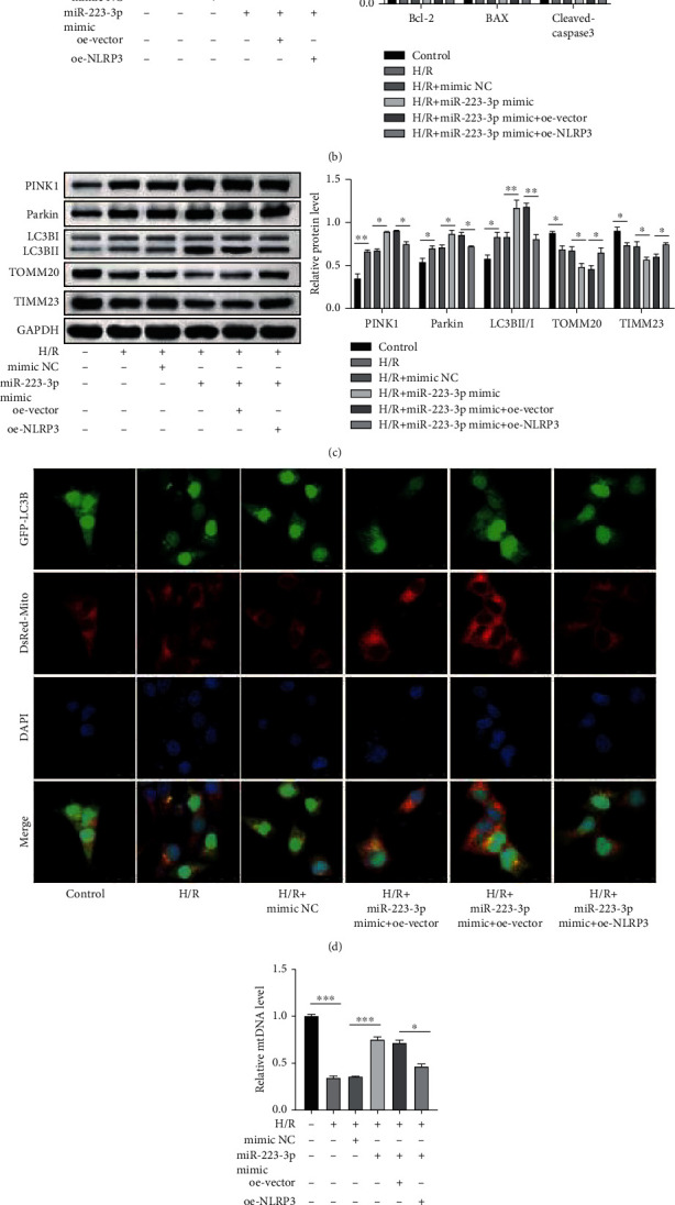
Figure 7.

miR-223-3p suppressed apoptosis and inflammasome activation and promoted mitophagy in H/R-induced TCMK-1 cells by targeting NLRP3. TCMK-1 cells were subjected to H/R stimulation and miR-223-3p-depleted exosome treatment. (a) H/R-induced cell apoptosis was determined through flow cytometry. (b) The protein levels of BAX, cleaved caspase-3, and Bcl-2 were confirmed by performing the western blot analysis. (c) Expressions of the mitophagy markers were detected using western blot analysis. (d) Mitophagosome formation was assessed based on the colocalization of the autophagy marker GFP-LC3B and the mitochondria marker Mito Tracker (DsRed-Mito). (e) The levels of mitochondrial DNA (mtDNA) were determined through qPCR. (f) The expressions of NLRP3, ASC1, and cleaved caspase 1 were determined using the western blot analysis. N = 3. ∗P < 0.05; ∗∗P < 0.01; ∗∗∗P < 0.001.
