Fig. 7.
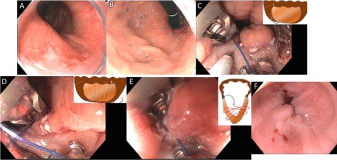
Resection and plication (RAP) technique for GERD in a patient after gastric bypass. a Endoscopic appearance of a wide open, patulous GEJ above the gastric pouch. b Retroflex view from the gastric pouch showing open hiatus, Hill Grade II-III. c After completion of piecemeal endoscopic mucosal resection (EMR) using a band ligation technique, the exposed muscularis propria is seen and suturing begins with the first bite taken distally at 8 o’clock position. d Bites are taken from 8 o’clock to 3 o’clock, alternating in a zigzag pattern (see inset) between distal and proximal sides of the mucosectomy area. The seventh bite seen here is at 4 o’clock at distal edge. e After the first running suture is cinched down, a second reinforcing suture is placed in a “V” formation. f Post-RAP appearance of the GEJ which is now much narrower with the creation of a short flap valve

