Figure 7.
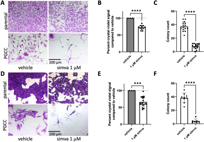
Simvastatin alone inhibits PGCC division. (A) 10X images of parental PPC1 cells or PGCC treated with DMSO or simvastatin. Daughter cells are shown mid-extrusion from DMSO treated PGCC while simvastatin treated PGCC lack progeny. (B) Growth inhibition in parental PPC1 exposed to simvastatin is quantified by the crystal violet viability assay, with DMSO treatment set at 100% viable. (C) Colony counts of filtered PGCC are quantified, with at least three independent experiments represented. (D) 10X images of parental MEL624 cells or their PGCC treated with DMSO or simvastatin. Statin-treated parental cells show slight growth inhibition as quantified in (E). Statin-treated PGCC show a loss of colony formation as quantified in (F), representing at least three independent experiments. ***p < 0.001, ****p < 0.0001.

