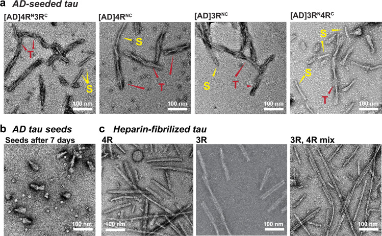
Fig. 2. Ultrastructural morphology of AD-tau seeded tau fibrils is distinct from that of heparin-fibrillized tau.
a TEM images of AD-tau seeded fibrils containing a 1:1 mixture of 15N-labeled 4R tau and 13C-labeled 3R tau, 4R-only tau, 3R-only tau, and a 1:1 mixture of 15N-labeled 3R tau and 13C-labeled 4R tau. All samples show predominantly twisted (T) fibrils whose widths alternate between ~10 nm and ~25 nm, similar to the paired helical filaments of AD tau. In addition, a minority of straight (S) fibrils are observed, which are similar to the straight filaments of AD tau. b AD tau seeds after 7 days of incubation without added recombinant monomers. The seeds are fivefold shorter than the amplified tau fibrils. No long fibrils are observed. c Heparin-assembled recombinant tau fibrils. These fibrils are nearly exclusively straight, and are longer and more uniform than the straight filaments in AD-tau seeded filaments. At least five different images from different regions of each grid were obtained for each sample. A side-by-side comparison of these fibrils can be found in Supplementary Fig. 2.

