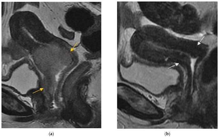
Figure 1.
(a) Sagittal FSE T2-weighted image shows soft tissue with high signal intensity consistent with a cervical tumor extended to the vagina (arrows). (b) Sagittal FSE T2-weighted image shows reconstitution of the normal T2-weighted hypointense cervical stroma (arrows), with disappearance of the tumoral mass after chemoradiation therapy.

