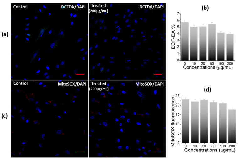
Figure 6.
Impact of nanosheets treatment on oxidative stress in hWJ-MSCs. (a) Confocal images, (b) Quantitative estimation of DCF-DA stained control and treated cells (200 µg/mL), showing no oxidative stress, (c) Confocal images, and (d) Quantitative estimation of MitoSOX-stained control and treated cells, indicating non-significant generation of mitochondrial superoxide in treated cells (scale bar: 50 µm).

