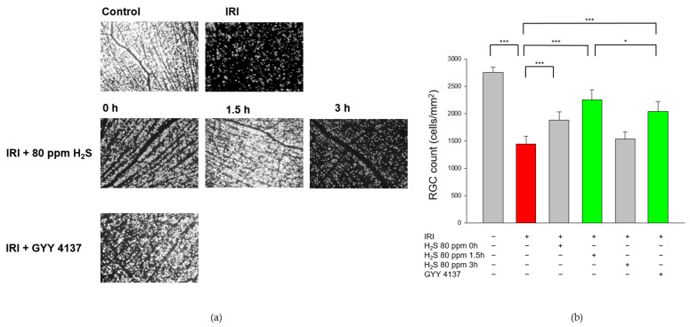
Figure 1.
Effect of H2S on retinal ganglion cell count after ischemia-reperfusion injury (IRI). Rats were subjected to unilateral retinal IRI and subsequently either received inhalative therapy with 80 ppm H2S directly and 1.5 h after IRI or an intravenous application of the slow-releasing H2S donor GYY 4137 immediately following IRI. (a) Representative flat mound images of fluoroscope-labeled retinal ganglion cells 7 days after IRI and respective treatment. (b) Quantification of retinal ganglion cell density [cells/mm2, data are mean ± SD, *** = p < 0.001, untreated vs. IRI (n = 8), IRI vs. IRI + 80 ppm H2S at 0 h (n = 8), vs. IRI + 80 ppm H2S at 1.5 h (n = 8), and vs. IRI + GYY 4137 (n = 6)]. The total numbers of surviving ganglion cells after inhalation of H2S with a delay of 1.5 h was significantly increased compared to intravenous treatment with GYY 4137 [* = p < 0.05, IRI + 80 ppm H2S at 1.5 h (n = 8) vs. IRI + GYY 4137 (n = 6)]. The red column illustrates the reduction of RGC by IRI, and the green columns highlight the positive effect of H2S and GYY.

