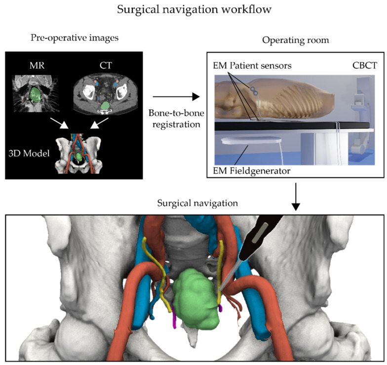
Figure 1.
Surgical navigation workflow showing live patient and pointer tracking by an electromagnetic (EM) navigation system. (Top left) Prior to surgery, a 3D model of the pelvic area is made by delineating all relevant structures on a preoperative CT and/or MRI scan. (Top right) During surgery, the procedure starts by placing EM patient sensors on the skin and imaging the patient’s position on the surgical table using a cone beam CT (CBCT). This image is then subsequently registered to the preoperative CT scan and linked with the 3D model. (Bottom) This registration process permits following the movements of the surgeon within the detailed 3D model via the use of an in vivo EM tracked pointer (black pointer).

