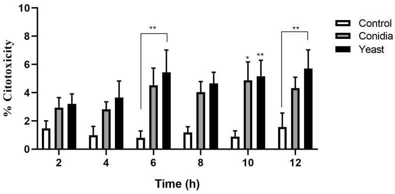
Figure 1.
Cytotoxicity of keratinocytes during infection by conidia and yeast cells of S. schenckii. Cell infection kinetics were performed with conidia and yeast cells of S. schenckii in keratinocytes with a 1:1 MOI, for 12 h. At each post-infection time, the cell culture supernatants were recovered. The assessment was performed with the CytoTox 96® Assay commercial kit using lactate dehydrogenase (LDH) detection. The data are presented as the mean ± standard deviation (SD) of three independent experiments. * p < 0.05, ** p < 0.005.

