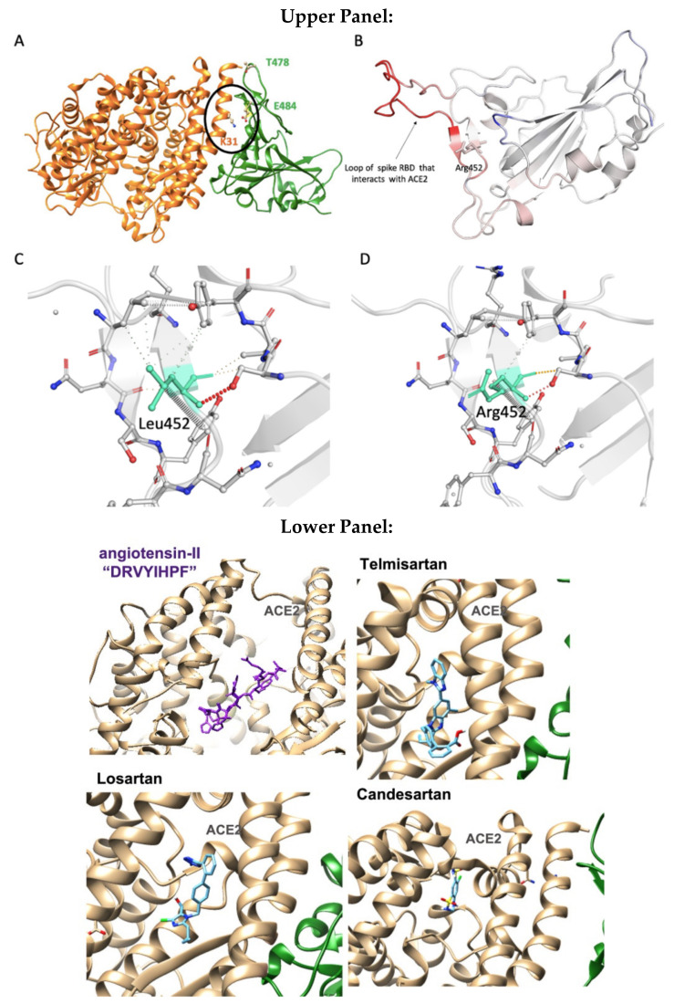
Figure 13.
Upper Panel: (A) Contacts between Lys31(ACE2) and Glu484 (S-protein RBD) in the human-virus interface. (B) Amino acids of S-protein RBD are colored according to the vibrational entropy change upon L452R mutation: blue represents a rigidification of the structure and red: a gain in flexibility. (C,D). Wild-type (L452) and mutant residue (R452) are colored in light-green and represented as sticks alongside with the surrounding residues which are involved on any type of interactions. Lower Panel: The binding site of AngII (purple), telmisartan, losartan, candesartan in the ACE2 zinc-domain groove (light gold) complexed with S-protein RBD (green).

