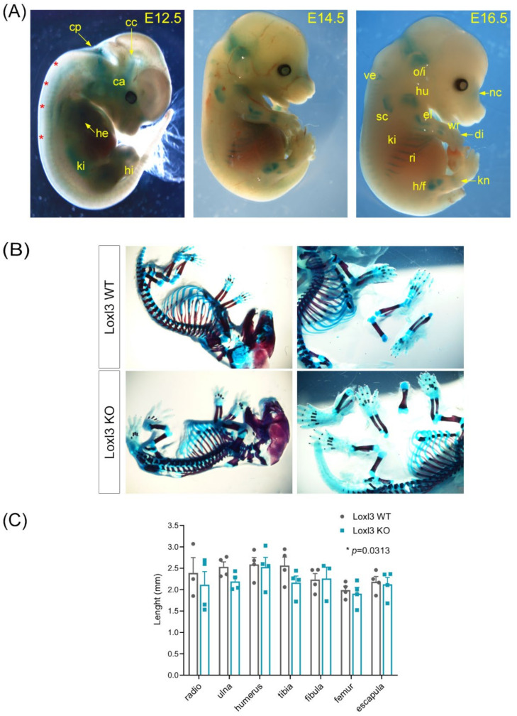
Figure 2.
Loxl3 expression during embryogenesis is associated with mouse skeletal development. (A) Loxl3 expression pattern in embryos at indicated developmental stages as depicted by X-Gal staining. Red asterisks indicate axial staining; cp, choroid plexus; cc, chondrocranium; ca, cochlear area; he, heart; ki, kidney; hi, hindlimbs; o/i, occipital and interparietal cartilaginous precursors; ve, vertebrae; hu, humerus; nc, nasal cartilages; el, elbow; sc, scapula; wr, wrist; di, digits; ri, ribs; s/f, hip/femur; kn, knee. (B) Representative images of Loxl3 WT (Loxl3+/+) and KO (Loxl3LacZ/LacZ) E18.5 embryos stained with alcian blue (cartilage)/alizarin red (bone) used to measure mineralised tissues. (C) Length (mm) of indicated bones from Loxl3 WT (Loxl3+/+) and KO (Loxl3LacZ/LacZ) embryos (E18.5). Individual values and mean with SEM from WT (n = 4) and KO (n = 4) littermate embryos are shown. p value was calculated by Wilcoxon matched-pairs signed rank test.

