Figure 3.
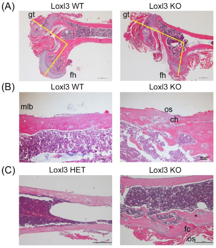
Embryonic loss of Loxl3 promotes abnormal skeletal development. (A) Images showing femoral head dysplasia found in several Loxl3 KO (Loxl3LacZ/LacZ) mice with atrophy of the femoral head (fh), hypertrophy of the greater trochanter (gt) and an abnormal angle between the fh and gt, displayed by yellow lines. (B) The lamellar diaphysis bone in Loxl3 KO (Loxl3LacZ/LacZ) animals is interrupted by woven bone, including few chondrocytes (ch) and active osteoblasts (os) in the periphery compared to the mature lamellar bone (mlb) in WT mice. (C) The diaphysis from Loxl3 KO (Loxl3LacZ/LacZ) femur displays a fibro-cartilaginous (fc) area with local deformation and bone marrow disorganisation not present in a littermate heterozygous mouse (HET, Loxl3+/LacZ). The asterisk depicts a connective channel between the periphery and deep inter-trabecular spaces. Scale bars, 100 µm.

